
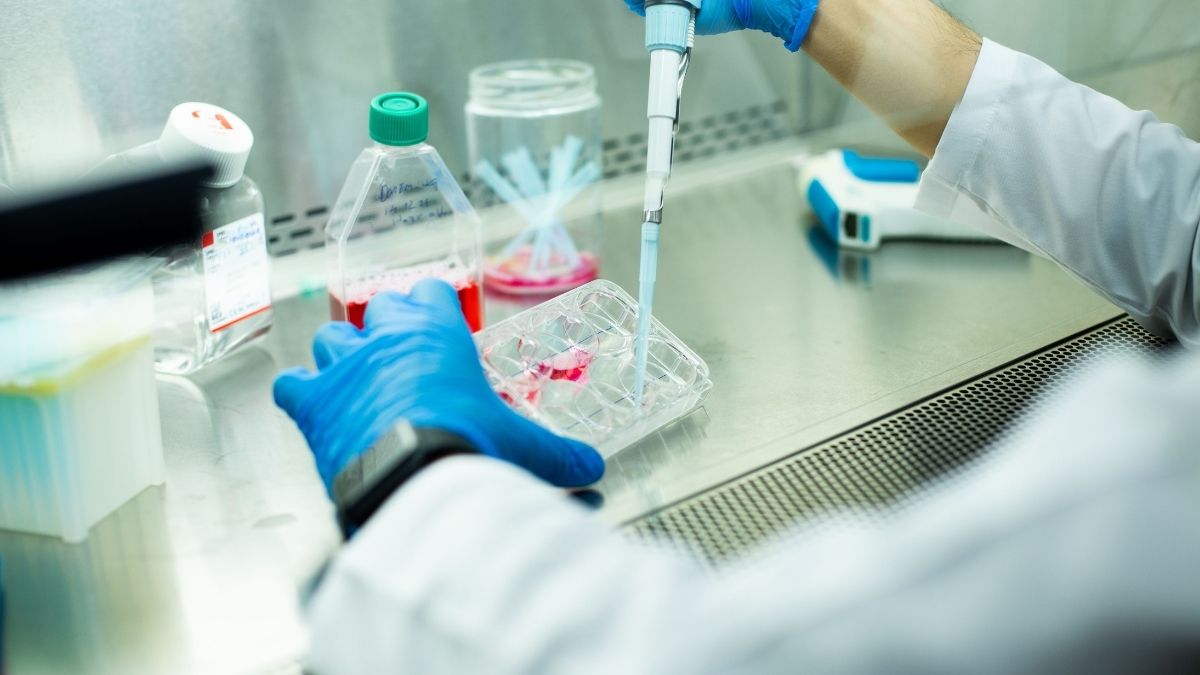
When we think about calcium, our minds often jump straight to bone health or the glass of milk we had with breakfast. While those are certainly important, there is a much more intricate story happening at the microscopic level. Inside every cell in the human body, calcium ions act as one of the most versatile and critical messengers. They dictate when a muscle should contract, when a neuron should fire, and even when a cell should begin the process of division or programmed death. Because this element is so central to life, scientists have spent decades perfecting ways to measure it. This is where the calcium assay comes into play, serving as a window into the otherwise invisible world of cellular communication.
The beauty of a calcium assay lies in its ability to translate a chemical event into a visual signal. By using specialised sensors or dyes that react to the presence of calcium ions, researchers can watch in real-time as signals ripple through a cluster of cells. This isn’t just academic curiosity; it is a fundamental part of how we develop new medicines and understand complex diseases. Whether it is investigating how the heart beats or how the brain processes information, tracking calcium flux is often the first step in solving the puzzle.

How the calcium assay works in the laboratory
At its core, a calcium assay is designed to detect changes in the concentration of intracellular calcium. In a resting cell, the level of calcium in the cytoplasm is kept extremely low compared to the environment outside the cell. When a cell is stimulated, calcium channels open, allowing a sudden influx of ions. To capture this moment, scientists use various types of indicators that change their light-emitting properties when they bind to calcium.
There are several common ways to set up these experiments, depending on the specific questions being asked:
- Fluorescent Indicators: These are chemical dyes that are loaded into the cells. When the cells are hit with a specific wavelength of light, the dyes glow. If calcium is present, the intensity or colour of that glow changes, allowing researchers to quantify the amount of calcium flux.
- Bioluminescent Proteins: Inspired by organisms like jellyfish, these proteins emit light naturally when they encounter calcium. This method is often preferred for long-term monitoring because it doesn’t require an external light source that might damage the cells.
- Genetically Encoded Indicators: For more advanced studies, researchers can engineer cells to produce their own calcium sensors. This allows for the study of specific types of cells within a complex tissue without affecting the surrounding environment.
The choice of method often depends on the speed of the biological process being studied. For example, heart cells beat very quickly, requiring a calcium assay that can capture rapid changes with high temporal resolution. On the other hand, studying the slow changes associated with cell growth might allow for a more gradual approach.
The critical role in cardiac research
Perhaps nowhere is the calcium assay more vital than in the study of the heart. Every single heartbeat is triggered by a massive, coordinated release of calcium within the cardiac muscle cells, a process known as excitation-contraction coupling. If this process is disrupted even slightly, it can lead to arrhythmias or heart failure. Researchers use assays to observe how heart cells—often derived from human stem cells—react to different stimuli or potential drugs.
By analysing the “calcium transient”—the rise and fall of calcium levels during a beat—scientists can determine if a heart cell is functioning normally. They look for specific characteristics in the data, such as:
- The peak height of the calcium release, which indicates the strength of the potential contraction.
- The rate of decay, showing how quickly the cell can clear calcium to prepare for the next beat.
- Irregularities or “sparks” that might suggest a risk of dangerous heart rhythms.
This level of detail is essential for safety pharmacology. Before a new drug can be approved for human use, it must be screened to ensure it doesn’t interfere with these delicate calcium signals. Using a high-quality calcium assay allows developers to identify potential cardiac side effects much earlier in the drug discovery process, saving time and, more importantly, protecting patient safety.
Advancing drug discovery through automation
In the past, conducting a calcium assay was a labour-intensive process that required a scientist to sit at a microscope and manually record data from one cell at a time. While this provided great detail, it wasn’t practical for testing thousands of different chemical compounds. Modern laboratories have transformed this through high-throughput screening (HTS).
Today, automated systems can run hundreds of assays simultaneously in multi-well plates. These systems are equipped with sophisticated cameras and software that can process massive amounts of data in minutes. This shift has revolutionised the way we approach diseases like diabetes, hypertension, and neurological disorders. By rapidly screening vast libraries of molecules, researchers can find “hits” that modify calcium signalling in a beneficial way, providing a starting point for new therapies.
However, automation brings its own set of challenges. Maintaining cell health across a large plate and ensuring that the dye is loaded evenly into every single cell requires a high degree of precision. This is why many pharmaceutical companies and academic institutions partner with specialised laboratories that have the expertise to optimise these protocols for consistency and accuracy.

The challenges of getting accurate data
While the technology has come a long way, performing a calcium assay is not without its hurdles. One of the primary concerns is the potential for the indicators themselves to interfere with the cell’s natural behaviour. Because fluorescent dyes bind to calcium, they can act as “buffers,” effectively soaking up the very ions they are supposed to be measuring. If the concentration of the dye is too high, it might dampen the signal or slow down the biological response.
Another issue is background noise. Cells naturally contain molecules that fluoresce, and the growth media used in the lab can also create light interference. Distinguishing the true calcium signal from this background clutter requires careful calibration and the use of sophisticated controls. Researchers must also account for “photobleaching,” where the fluorescent signal fades over time due to exposure to light, which can make long-term experiments difficult to interpret.
To overcome these obstacles, the industry is moving towards ratiometric imaging. This technique uses dyes that shift their emission wavelength when bound to calcium. By taking the ratio of the signals at two different wavelengths, scientists can cancel out many of the variables, such as differences in dye concentration or cell thickness, leading to much more reliable and reproducible data.
Looking toward the future of cellular imaging
As we look forward, the evolution of the calcium assay is likely to be driven by improvements in imaging speed and the integration of artificial intelligence. We are already seeing the emergence of 3D organoid models—miniature versions of organs grown in the lab—which provide a more realistic environment for testing than traditional flat cell cultures. Measuring calcium flux in these complex 3D structures requires even more advanced optical techniques and data processing power.
Furthermore, the combination of calcium imaging with other modalities, such as electrophysiology, is providing a more holistic view of cell function. Instead of just looking at one piece of the puzzle, researchers can now see how electrical changes and chemical signals work together in real-time. This integrated approach is paving the way for personalised medicine, where a patient’s own cells could be used in a calcium assay to determine which specific medication will be most effective for their unique genetic makeup. The precision of these measurements continues to refine our understanding of biology at its most fundamental level.

Elli specializes in leadership development, organisational change, and strategic decision-making to enhance business performance.
